Back to Profile Page

Tree of Life Media Contributed By Dirk Redecker

ID Thumbnail Media Data
7348
Comments Arbuscule within a root cell, stained with Chlorazol Black E.
Size Arbuscule diameter is approximately 19 µm
Copyright © Kerstin Wex
Image Use restricted
Attached to Group Glomeromycota: view page image collection
Title arbuscule.jpg
Image Type Photograph
Image Content Specimen(s)
ID 7348
7349
Scientific Name Plantago media, Glomus clarum
Comments Roots of Plantago media (plantain) colonized with Glomus clarum. Note the hyphae and spores on the roots.
Size Diameter of larger spores is approximately 200 µm
Copyright © Dirk Redecker
Image Use restricted
Attached to Group Plantaginaceae (Lamiales): view page image collection
Glomeromycota: view page image collection
Title lclr_myc.jpg
Image Type Photograph
Image Content Specimen(s)
ID 7349
7350
Scientific Name Glomus sp. S328
Comments Spore of Glomus sp. S328 with attached hypha.
Size Spore diameter is approximately 80 µm.
Copyright © 2000 American Association for the Advancement of Science
Image Use restricted
Attached to Group Glomeromycota: view page image collection
Title s328_small.jpg
Image Type Photograph
Image Content Specimen(s)
ID 7350
7370
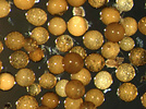
Scientific Name Scutellospora castanea
Comments Spores of Scutellospora castanea
Life Cycle Stage spore
Size Spore diameter approximately 0.22 mm
Copyright © Annemarie Brennwald
Image Use restricted
Attached to Group Scutellospora (Diversisporales): view page image collection
Title scutellospora_sm.jpg
Image Type Photograph
Image Content Specimen(s)
ID 7370
7372
Scientific Name Glomus sinuosum
Comments Section of a sporocarp of Glomus sinuosum (formerly Sclerocystis sinuosa). Spores are arranged around a center of interwoven hyphae and covered by a "peridium".
Acknowledgements Isolate courtesy of INVAM
Copyright © Dirk Redecker
Image Use restricted
Attached to Group Glomeromycota: view page image collection
Title sinuosum_adj2.jpg
Image Type Photograph
ID 7372
7373
Comments Fossil glomalean spore
Specimen Condition Fossil -- Period: Ordovician of Wisconsin
Size size bar 0.05 mm
Copyright © 2000 American Association for the Advancement of Science
Image Use restricted
Attached to Group Glomeromycota: view page image collection
Title fossilspore2.jpg
Image Type Photograph
Image Content Specimen(s)
ID 7373
7374
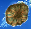
Scientific Name Archaeospora leptoticha
Comments Spore of Archaeospora leptoticha cracked open under a microscope cover slide. The components of spore wall structure became visible, including a richly ornamented wall.
Acknowledgements Isolate courtesy of INVAM
Life Cycle Stage spore
Size Uncracked spore diameter approximately 0.02 mm
Copyright © Dirk Redecker
Image Use restricted
Attached to Group Archaeospora (Archaeosporales): view page image collection
Title agrd_bw1.jpg
Image Type Photograph
Image Content Specimen(s)
ID 7374
Please note: Most images and other media displayed on the Tree of Life web site are protected by copyright, and the ToL cannot act as an agent for their distribution. If you would like to use any of these materials for your own projects, you need to ask the copyright owner(s) for permission. For additional information, please refer to the ToL Copyright Policies.

Contribute to the ToL

ToL Contributions

Ways to Contribute

Use of Contributions

Scientific Content

Building Treehouses

Images/Media

  go to the Tree of Life home page
top