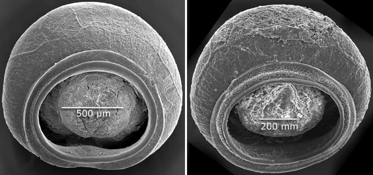
Suckers of two of paratypes were examined with the scanning electron microscope (SEM). Suckers were prepared by dehydration in absolute ethanol then air drying.
- Arm suckers
- Tentacular club suckers


Figure. Oral view of arm suckers of Histioteuthis sp. A, 40 mm ML, Hawaiian waters (21°20'N, 158°20'W). Left - Arm II, sucker V8 (sucker 8 of ventral series). Right - Arm III, sucker V4. Photographs by R. Young.


Figure. Oral view of arm IV sucker V4 of Histioteuthis sp. A, same specimen as above. Photograph by R. Young.


Figure. Oral views of club suckers of Histioteuthis sp. A, same specimen as above, from the two ventral-most series on the club manus. Their positions on the club are indicated on the photograph below. The smaller sucker to the left is from the submarginal series and the sucker to the right is from the marginal series. Photographs by R. Young.


Figure. Oral view of the club of Histioteuthis sp. A from which the suckers for the adjacent SEM photographs were taken. The bottom circle indicates the location of the two suckers viewed above. The two upper circles indicate the location of the suckers viewed below. Suckers from the two marginal series had been removed prior to taking the photograph. Photograph by R. Young.


Figure. Oral views of club suckers from the central region of the manus of Histioteuthis sp. A, same specimen as previous photographs. Their positions on the club are indicated on the photograph above.. Photographs by R. Young.


Figure. Oral view of sucker from ventral-marginal series of the club of Histioteuthis sp. A, 41 mm ML, same capture locality as the previous specimen. Left - oral view of the club with the sucker encircled that was examined with SEM. Right - SEM photograph of the encircled sucker. Sucker was damaged distally during preparation. Photographs by R. Young.


 Go to quick links
Go to quick search
Go to navigation for this section of the ToL site
Go to detailed links for the ToL site