Abralia trigonura
Kotaro Tsuchiya and Richard E. YoungIntroduction
A. trigonura belongs to the Heterabralia subgenus. It is characterized by having two or three hooks on the manus, the left arm hectocotylized, and complex eye photophores. This species resembles A. andamanica but the latter is separable by its robust tail and dark body color.
Characteristics
- Tentacle clubs
- Two or three hooks on ventral side.
- Two rows of large suckers on dorsal side of manus.
- Hectocotylus
- Left ventral arm of male hectocotylized.
- Hectocotylus with two different sized off-set flaps.
- Suckers
- Suckers dentate all aroung.
- Head
- Eye Photophores: Five, complex organs: two large terminal opaque organs and three intermediate silvery organs.
- Beaks: Descriptions can be found here: Lower beak; upper beak.
- Integumental Photophores
- Ventral mantle and head with scattered arrangement of integumental organs.
- Ventral mantle and head with scattered arrangement of integumental organs.
- Mantle apex ("tail")
- Broad and extends well beyond conus of gladius.
- Broad and extends well beyond conus of gladius.
- Reproductive structures
 Click on an image to view larger version & data in a new window
Figure. Dorsal view of the receptacle for spermatangia in female A. trigonura. Anterior is left. Left side - Note the nuchal cartilage and collar on either side. Middle - Deep pit that holds the spermatangia. Right - Mantle and gladius folded posteriorly showinng the receptacle located where the mantle and visceral sac fuse. Drawing from Burgess (1991, p. 115, Fig. 1K)


Figure. Ocular photophores of A. trigonura. Left - Ventral view. Drawing from Tsuchiya (2000). Right - Postero-ventral view. Photograph by R. Young of a fresh squid with overlying skin removed.
Comments
A. trigonura is similar to A. andamanica, A. siedleckyi, A. heminuchalis, and A. veranyi in its complex ocular photophores and left ventral arm hectocotylization.
Life history
Age and reproduction
Female longevity is up to 6 months, and sexual maturity is reached at 3.5 months. Male longevity is the same as female, and sexual maturity is reached at 2.5 months. The smallest mature female is 31mm DML, and 80% of females are mature at 35mm DML. Males sexually mature between 23-27mm DML. This species seems to be a multiple spawner, and females spawn eggs every few days. Female batch fecundity was roughly 290-430 eggs (Young and Mangold, 1994).
Eggs
Spawned eggs are slightly ovoidal, 0.9 mm x 0.79 mm, usually with a slightly greenish tint, clear, and with a sticky colorless jelly coating (Young and Harman, 1985).


Figure. Left - "Egg" (actually early embryo) of A. trigonura taken from a plankton tow off Hawaii. The egg is surrounded by a gelationus oviducal secretion with debris attached. Right - Newly hatched paralarva of A. trigonura. Photographs by R. Young.
Paralarvae
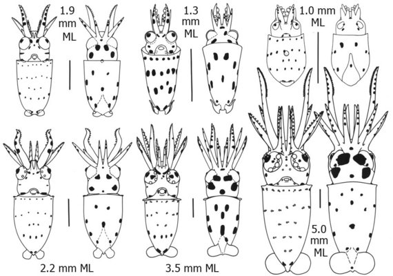
The distinctive chromatophore patterns, especially that of the ventral mantle, can be followed from hatching to the largest paralarva illustrated (5.0 mm ML) where each ventral mantle chromatophore is accompanied by a photophore. That is, the initial chromatophore pattern is duplicated with photophores as the paralarva grows.
Distribution
Vertical Distribution
In the Hawaiian waters, this species was considered to be an important component of the mesopelagic boundary community (Reid et al., 1991), and vertically migrates from upper mesopelagic depths during the day to the upper 50 m at night (Young, 1978). It generally stays during the daytime >5m above the ocean floor (Young, 1995). Young stages occur primarily outside the mesopelagic boundary zone. Adults inhabit the outer mesopelagic boundary zone during daytime, and migrate to the inner mesopelagic boundary zone during night (Young, 1995).
Geographical Distribution
This species is broadly distributed in tropical West to Central North Pacific (Hidaka and Kubodera, 2000).
References
Hidaka, K. and T. Kubodera. 2000. Squids of the genus Abralia (Cephalopoda: Enoploteuthidae) from the western tropical Pacific with a description of Abralia omiae, a new species. Bull. Mar. Sci. 66: 417-443.
Tsuchiya, K. 2000. Illustrated book of the Enoploteuthidae. In: Okutani T., ed. True face of Watasenia scintillans. Tokai University Press, Tokyo, p 196–269. (in Japanese)
Young, R.E. 1995. Aspects of the natural history of pelagic cephalopods of the Hawaiian mesopelagic-boundary region. Pacific Science, 49:143-155.
Young, R.E. and Harman, R.F. 1985. Early life history stages of enoploteuthin squids (Cephalopoda: Teuthoidea: Enoploteuthidae) from Hawaiian waters. Vie Milieu, 35:181-201.
Young, R.E. and Mangold, K.M. 1994. Growth and reproduction in the mesopelagic boundary squid Abralia trigonura. Marine Biology, 119: 413-421.
Title Illustrations

| Scientific Name | Abralia trigonura |
|---|---|
| Location | off Hawaii |
| Specimen Condition | Live Specimen |
| Life Cycle Stage | adult |
| View | lateral |
| Image Use |
 This media file is licensed under the Creative Commons Attribution-NonCommercial License - Version 3.0. This media file is licensed under the Creative Commons Attribution-NonCommercial License - Version 3.0.
|
| Copyright |
© 2000

|
About This Page
Figures from Burgess (1991) are printed with the Permission of the Bulletin of Marine Science.

Tokyo University of Fisheries, Tokyo, Japan

University of Hawaii, Honolulu, HI, USA
Page copyright © 2018 and
 Page: Tree of Life
Abralia trigonura .
Authored by
Kotaro Tsuchiya and Richard E. Young.
The TEXT of this page is licensed under the
Creative Commons Attribution-NonCommercial License - Version 3.0. Note that images and other media
featured on this page are each governed by their own license, and they may or may not be available
for reuse. Click on an image or a media link to access the media data window, which provides the
relevant licensing information. For the general terms and conditions of ToL material reuse and
redistribution, please see the Tree of Life Copyright
Policies.
- Content changed 20 February 2018
Citing this page:
Tsuchiya, Kotaro and Richard E. Young. 2018. Abralia trigonura . Version 20 February 2018 (under construction). http://tolweb.org/Abralia_trigonura/19657/2018.02.20 in The Tree of Life Web Project, http://tolweb.org/


 Go to quick links
Go to quick search
Go to navigation for this section of the ToL site
Go to detailed links for the ToL site